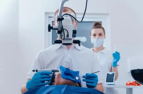
The process begins with a thorough diagnosis, typically involving X-rays and clinical evaluation, to assess the extent of the issue. Once confirmed, the dentist administers local anesthesia to ensure the patient’s comfort. A small access opening is then created to reach the pulp chamber and root canals. Using specialized instruments and magnification tools, the infected or damaged pulp is delicately removed. The canals are meticulously cleaned, disinfected, and shaped to eliminate any remaining debris or bacteria. After this, the canals are filled with a biocompatible material, often gutta-percha, to seal the space and prevent further infection.
Microscopic root canal treatment
The process begins with a thorough diagnosis, typically involving X-rays and clinical evaluation, to assess the extent of the issue. Once confirmed, the dentist administers local anesthesia to ensure the patient’s comfort. A small access opening is then created to reach the pulp chamber and root canals.
Using specialized instruments and magnification tools, the infected or damaged pulp is delicately removed. The canals are meticulously cleaned, disinfected, and shaped to eliminate any remaining debris or bacteria. After this, the canals are filled with a biocompatible material, often gutta-percha, to seal the space and prevent further infection.